3D Surgical Atlases of the Murine Head


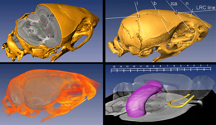
Provided below for download are high resolution surgical MRI / CT atlases of the adult murine head for strains C57Bl/6j and 129S1/SvIMJ.   3D neuroanatmical labels are also provided.   In the event that these files cannot be natively read, several links to file readers are provided.   Papers provided below discuss stereotactic alignment and labelling considerations as applied across different murine strains; as well as details of our variational / probabilistic MRI atlases of the adult mouse brain for strains CD1, C57Bl/6j and 129S1/SvIMJ. A map of image file conventions and file inter-operability among common freeware programs can be seen here. Copies of these freeware programs can be obtained on the following page under the "3D imaging" section. The surgical atlases below are part of the NeuroMouse project.


   3D atlas: C57Bl/6J brain   (13.5 MB)

   3D atlas: C57Bl/6J (labels)   (6.8 MB)

   3D atlas: C57Bl/6J (dorsal skull)   (6.7 MB)

   3D atlas: C57Bl/6J (complete skull)   (16 MB)


   3D atlas: 129Sv/Imj (brain)   (13.5 MB)

   3D atlas: 129Sv/Imj (labels)   (6.8 MB)

   3D atlas: 129Sv/Imj (dorsal skull)   (6.7 MB)

   3D atlas: C57Bl/6J (complete skull)   (16 MB)


     MNC file reader: OCCI Viewer Viewer file.zip   (19.7 MB)

     MNC file reader:  Display and Register
     Display:  Setup Guide
     Display:  Software Guide
     Register:   Software Guide

     MRI file reader: MRIcro download
     MRIcro Software Guide


Primary atlas citation:

  • Chan E., Kovacevic N., Ho S.K.Y., Henkelman R.M., and Henderson J.T. “Development of a high resolution three dimensional surgical atlas of the murine head for strains 129S1/SvImJ and C57Bl/6J using magnetic resonance imaging and micro-computed tomography and micro-CT”, Neuroscience, Volume 144(2), 604-615, 2007 (Epub Nov. 13, 2006).

  • Additional atlas papers:

  • Chen X.J., Kovacevic N., Lobaugh N.J., Sled J.G., Henkelman R.M., and Henderson J.T. “Neuroanatomical differences between mouse strains as shown by high-resolution 3D MRI”, Neuroimage 29(1): 99-105, 2006 (Epub Aug. 9, 2005).
  • Kovacevic N., Henderson J.T., Chan E., Lifshitz N., Bishop J., Evans A.C., Henkelman R.M., Chen X.J. “A Three-dimensional MRI Atlas of the Mouse Brain with Estimates of the Average and Variability”, Cerebral Cortex, 15(5):639-45, 2005 (Epub Sept. 1, 2004)
  • Kovacevic N., Chen X.J., Sled J., Henderson J.T., and Henkelman R.M “Deformation-based representations of groupwise average and variability”, Medical Image Computing and Computer-Assisted Intervention (MICCAI 7th), ISBN 3-540-20464-4, vol. 3216, 615-622, 2004.


  • Imaging Links:


    Principle open source medical imaging file types:

    Example: Multipage TIFF (.tif, .tiff, 12MB)

    Principle medical sub-formats:

    Example: Dicom (.dcm - 9.2MB) | Example: MINC (.mnc/.mnc2 - 13.5MB)

    Example: Analyze.hdr (1k) img (54.5MB)


    Local Imaging Sites:

    Murine Imaging and Histology (MIH) Core | Software / Imaging Standards

    Toronto Center for Phenogenomics (TCP) | U of T Program in Neuroscience


    Imaging Atlases:

    Scalable Brain Atlas: Allen Mouse Brain volumetric atlas 2012

    Scalable Brain Atlas: Waxholm Space delineation 2012

    The e-Mouse Atlas Project (EMAP)

    High res. histologic atlas:     Brain Maps.org

    Histologic atlas:     MBL mouse brain atlas

    Histologic atlas:     Harvard mouse brain atlas


         MIH Homepage

         Henderson Laboratory Home Page

          Send mail to Dr. Henderson: jeff.henderson@utoronto.ca